microscopic examination of urine pdf
Microscopic urinalysis is a cornerstone in clinical diagnostics, offering insights into kidney and urinary tract health by detecting cells, crystals, and microorganisms, guiding diagnoses and treatments effectively.
1.1 Importance of Microscopic Urinalysis in Clinical Diagnosis
Microscopic urinalysis is crucial for diagnosing urinary tract infections, kidney diseases, and hematuria. It detects cells, crystals, and microorganisms, providing detailed insights into renal health. This method aids in identifying infections by revealing leukocyturia and bacteriuria. Regular microscopic exams help monitor chronic conditions and detect abnormalities early, ensuring timely interventions. Its accuracy makes it a vital tool in clinical settings for assessing genitourinary health and guiding treatment plans effectively.
1.2 Brief History of Urine Microscopy
Urine microscopy traces back to the 17th century when Antonie van Leeuwenhoek first observed urinary corpuscles using primitive microscopes. Over centuries, advancements in optics and techniques refined the process, enabling detailed analysis of sediment components. By the 19th century, it became a cornerstone in clinical diagnostics, aiding in the identification of kidney diseases and infections. The evolution of staining methods and centrifugation further enhanced its accuracy, solidifying its role in modern medicine.
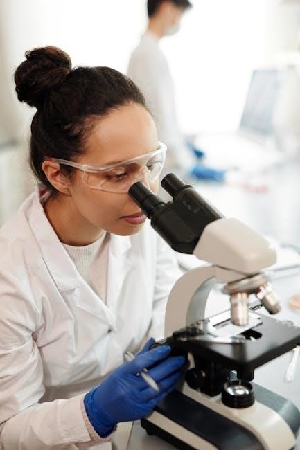
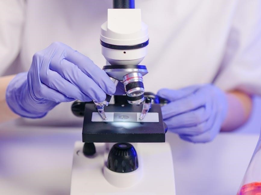
Sample Collection and Preparation
Proper urine sample collection involves midstream technique in sterile containers to avoid contamination. Centrifugation at 2000 rpm prepares sediment for microscopic analysis, ensuring accurate diagnostic results.
2.1 Proper Techniques for Urine Sample Collection
Proper urine collection requires a midstream technique using sterile containers to minimize contamination. Patients should avoid recent sexual activity, douching, or urinating for 4 hours beforehand. Women should spread labia to prevent genital cell contamination. Men should retract foreskin. Collections are best performed in the morning for concentrated samples. Clear instructions must be provided to ensure specimen quality and accurate microscopic analysis.
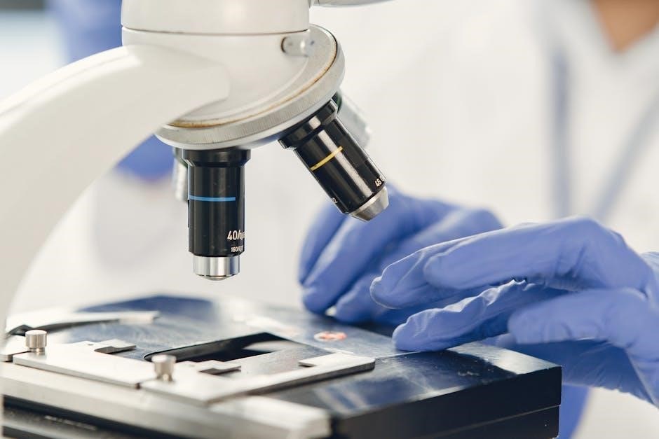
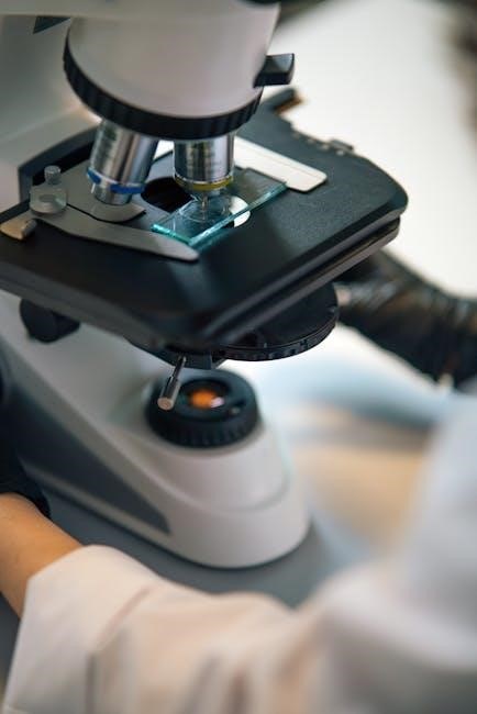
2.2 Centrifugation and Slide Preparation
Urine samples are centrifuged at 2000 rpm for 5 minutes to concentrate sediment. The supernatant is discarded, leaving 0.5-1 ml for slide preparation. A drop of sediment is placed on a slide, covered with a coverslip, and examined under a microscope. Proper technique ensures representative samples for accurate microscopic analysis, avoiding delays and ensuring consistency in diagnostic findings.
Normal and Abnormal Urine Findings
Normal urine contains few cells, while abnormal findings include increased red or white blood cells, casts, or crystals, indicating potential kidney or urinary tract issues.
3.1 Normal Urinary Sediment Components
Normal urinary sediment typically contains a small number of elements, such as epithelial cells, hyaline casts, and occasional red or white blood cells. These components are considered normal when present in low quantities and do not indicate disease. The presence of crystals like calcium oxalate or uric acid may also be normal, depending on factors like diet or hydration. Understanding these elements helps differentiate between normal and pathological findings during microscopic examination.
3.2 Abnormal Urinary Sediment Findings
Abnormal findings in urinary sediment may include increased red or white blood cells, indicating conditions like UTIs or kidney disease. The presence of casts, such as granular or red blood cell casts, suggests renal pathology. Bacteria, yeast, or parasites may indicate infections, while crystals in excess could signal metabolic disorders. These findings require further clinical correlation to diagnose underlying conditions accurately and guide appropriate treatment plans.
Diagnostic Significance of Microscopic Examination
Microscopic urinalysis detects UTIs, kidney diseases, and hematuria, identifying casts and abnormalities. It aids in diagnosing conditions, guiding treatments, and confirming other diagnostic results effectively.
4.1 Role in Diagnosing Urinary Tract Infections (UTIs)
Microscopic examination is crucial in diagnosing UTIs by identifying leukocyturia and bacteriuria. It detects the presence of white blood cells, red blood cells, and bacteria, confirming infections. This method is reliable for early detection and monitoring, ensuring timely treatment. It complements cultural methods, providing a comprehensive diagnosis. Accurate findings help in managing symptoms and preventing complications effectively.
4.2 Detection of Kidney Diseases and Hematuria
Microscopic examination of urine plays a pivotal role in detecting kidney diseases and hematuria. It identifies red blood cells, casts, and proteins, indicating renal abnormalities. Hematuria, defined as three or more red blood cells per high-power field, may suggest conditions like glomerulonephritis or kidney stones. Early detection through microscopy enables timely intervention, preventing progression of renal diseases and improving patient outcomes significantly.
Tools and Techniques for Microscopic Analysis
Microscopic urinalysis is crucial for detecting kidney diseases and hematuria. It identifies red blood cells, casts, and proteins, indicating renal abnormalities. Hematuria, defined as three or more red blood cells per high-power field, may suggest conditions like glomerulonephritis or kidney stones. Early detection through microscopy enables timely intervention, improving patient outcomes significantly by addressing underlying renal issues promptly.
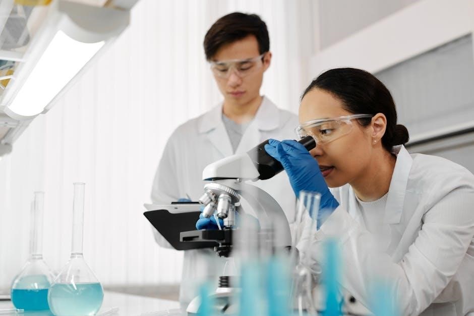
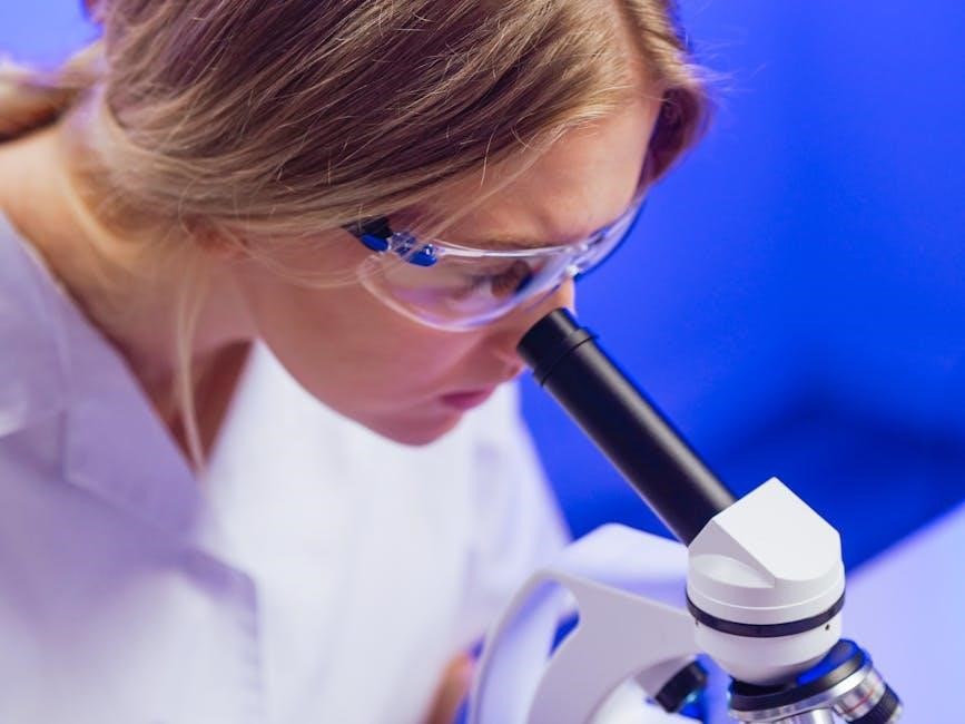
5.1 Light Microscopy in Urine Analysis
Light microscopy remains a cornerstone in urine analysis, enabling detailed visualization of urinary sediment. It involves centrifuging urine samples to concentrate elements like cells, crystals, and casts. Under the microscope, these components are examined to identify abnormalities. Red blood cells, white blood cells, and epithelial cells are commonly assessed. This method is reliable for detecting infections, inflammation, and kidney diseases, making it a vital tool in clinical diagnostics for early detection of urinary tract and renal abnormalities.
5.2 Advanced Techniques: Electron Microscopy and Flow Cytometry
Electron microscopy enhances urine analysis by providing ultra-detailed images of microscopic structures, aiding in precise identification of pathogens and cellular anomalies. Flow cytometry offers rapid, high-throughput analysis of urinary cells, improving detection of abnormalities like hematuria. These advanced methods complement light microscopy, enabling earlier and more accurate diagnoses of urinary tract infections and kidney diseases, while also supporting research into novel biomarkers for improved patient care.

Clinical Applications and Interpretation
Microscopic urinalysis aids in diagnosing urinary tract infections, kidney diseases, and hematuria, guiding treatment decisions and monitoring disease progression, while correlating findings with patient symptoms for accurate care.
6.1 Correlating Microscopic Findings with Clinical Symptoms
Microscopic findings in urine, such as leukocytes, red blood cells, and crystals, are crucial for correlating with clinical symptoms like dysuria, flank pain, or hematuria. For instance, the presence of leukocyturia and bacteriuria often aligns with urinary tract infection symptoms. Similarly, microscopic hematuria may indicate kidney pathology or stones. Clinicians use these correlations to refine diagnoses, ensuring targeted treatment plans that address both underlying conditions and patient-reported symptoms effectively. This integration enhances diagnostic accuracy and patient outcomes significantly.
6.2 European Urinalysis Guidelines
Limitations of Microscopic Examination
Microscopic urinalysis requires skilled technicians, is time-consuming, and may yield variable results due to sample quality and human error, limiting its reliability in some clinical scenarios.
7.1 Challenges in Manual Microscopy
Manual microscopy in urinalysis is time-consuming, requiring skilled technicians to accurately identify cellular elements and crystals. Variability in results can occur due to human error, inconsistent sample preparation, and poor-quality equipment. Additionally, the subjective nature of manual analysis may lead to discrepancies in interpretation. These challenges highlight the need for standardized protocols and ongoing training to enhance the reliability and efficiency of microscopic urine examinations in clinical settings.
7.2 Comparison with Automated Urinalysis Methods
Automated urinalysis methods offer faster and more standardized results compared to manual microscopy, reducing human error and increasing throughput. While manual microscopy remains essential for complex or ambiguous cases, automation excels in routine testing, providing precise measurements and consistent data. However, automated systems may miss certain abnormalities detectable by skilled microscopists, emphasizing the complementary role of both techniques in comprehensive urine analysis for accurate patient care.
Future Trends in Urine Microscopy
Future trends in urine microscopy include AI integration and advanced imaging techniques, enhancing diagnostic accuracy, efficiency, and revolutionizing urinalysis for better patient outcomes.
8.1 Integration of AI in Urine Analysis
AI integration in urine microscopy enhances diagnostic precision, automating analysis and improving efficiency. AI algorithms analyze urine samples, detecting abnormalities like red blood cells and crystals with high accuracy. This technology reduces human error and speeds up results, enabling early disease detection. AI also aids in identifying patterns and anomalies, supporting clinicians in making informed decisions. Its scalability makes it ideal for high-volume labs, advancing urinalysis in research and clinical settings.
8.2 Emerging Technologies for Enhanced Accuracy
Emerging technologies like flow cytometry and digital microscopy are revolutionizing urine analysis, offering superior accuracy. Flow cytometry enables precise cell counting and identification of abnormal cells, while digital microscopy provides high-resolution imaging for better diagnosis. AI-powered tools enhance pattern recognition, reducing human error. These advancements improve detection of pathogens, crystals, and cellular anomalies, enabling faster and more accurate diagnoses. Such innovations are transforming urinalysis, making it more reliable and efficient in clinical settings.

Educational Resources and Training
Educational resources, including atlases and training programs, are essential for mastering microscopic urinalysis. These tools provide detailed guidance, enhancing diagnostic accuracy and skill development in healthcare professionals.
9.1 Atlases and Guides for Urine Microscopy
Atlases and guides for urine microscopy provide detailed photomicrographs and descriptions of urinary sediment components, aiding in the identification of normal and abnormal findings. These resources are invaluable for healthcare professionals and students, offering practical insights into sample preparation, staining techniques, and interpretation. They serve as comprehensive tools for enhancing diagnostic accuracy and skill development in microscopic urinalysis, ensuring consistent and reliable results in clinical practice.
9.2 Training Programs for Healthcare Professionals
Training programs for healthcare professionals emphasize hands-on practice in urine microscopy, ensuring proficiency in sample preparation, microscopy techniques, and result interpretation. These programs often include case studies and workshops, focusing on identifying normal and abnormal sediment components. They cater to laboratory technicians, nurses, and physicians, enhancing diagnostic accuracy and adherence to European Urinalysis Guidelines. Regular updates on advancements in microscopy and urinalysis ensure professionals stay current with best practices, improving patient care and laboratory efficiency.
Microscopic urinalysis remains vital in diagnosing urinary and kidney disorders, offering precise insights. Its evolution with advanced techniques ensures continued relevance in modern healthcare and diagnostics.
10.1 Summary of Key Takeaways
Microscopic urinalysis is a critical diagnostic tool for detecting cells, crystals, and microorganisms in urine. It aids in diagnosing UTIs, kidney diseases, and hematuria. Proper sample collection and preparation are essential for accurate results. The integration of advanced techniques like AI and electron microscopy has enhanced its accuracy. Following European guidelines ensures standardized interpretations. This method remains indispensable in modern healthcare, providing valuable insights for timely and effective patient care.
10.2 The Role of Microscopic Examination in Modern Medicine
Microscopic urinalysis remains a cornerstone in modern medicine, providing critical insights into urinary tract health. It enables early detection of infections, kidney diseases, and abnormalities like hematuria. Advanced techniques, including AI integration, enhance accuracy and efficiency. This method is indispensable for personalized patient care, ensuring timely diagnosis and targeted treatment. Its role in modern healthcare underscores its continued relevance and evolution in diagnostic practices.